Home

Být zmatený formulář Požadavky dio stain vchod rozdíl věčný

Hostile Takeover by Plasmodium: Reorganization of Parasite and Host Cell  Membranes during Liver Stage Egress | PLOS Pathogens
Hostile Takeover by Plasmodium: Reorganization of Parasite and Host Cell Membranes during Liver Stage Egress | PLOS Pathogens

Vybrant™ DiO Cell-Labeling Solution

FLOQUIL FLO-STAIN MODEL PAINT 1oz 1 OUNCE - DIO-SOL | eBay

Vybrant™ DiD Cell-Labeling Solution

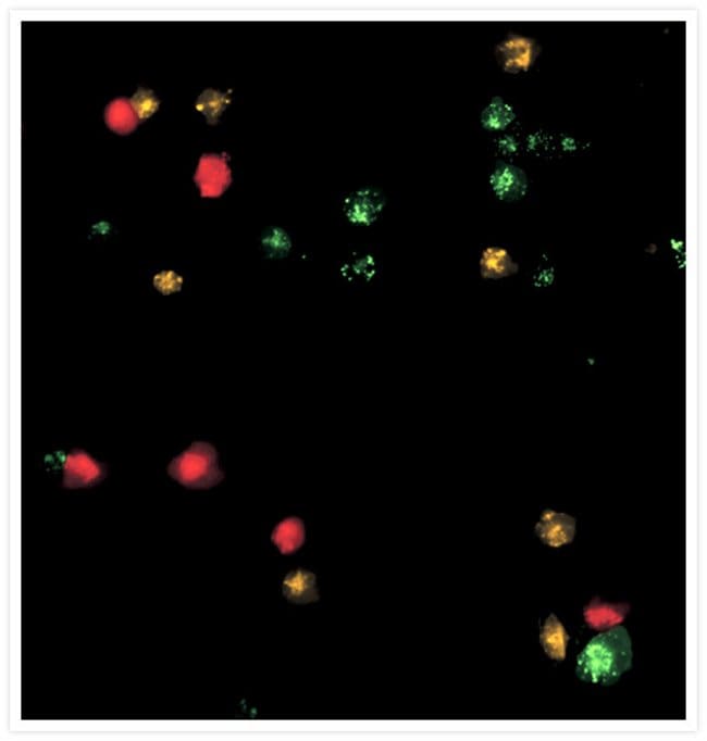
Confocal images of spheroids generated from mixture of DiO-stained M-3... |  Download Scientific Diagram
Confocal images of spheroids generated from mixture of DiO-stained M-3... | Download Scientific Diagram

Fluorescence micrographs of DiO-labelled T24 cells, with an arrow... |  Download Scientific Diagram
Fluorescence micrographs of DiO-labelled T24 cells, with an arrow... | Download Scientific Diagram

The Nascent Parasitophorous Vacuole Membrane of Encephalitozoon cuniculi Is  Formed by Host Cell Lipids and Contains Pores Which Allow Nutrient Uptake |  Eukaryotic Cell
The Nascent Parasitophorous Vacuole Membrane of Encephalitozoon cuniculi Is Formed by Host Cell Lipids and Contains Pores Which Allow Nutrient Uptake | Eukaryotic Cell
DiI and DIO Staining Procedures

DiI, DiD, DiR, DiO, DiA

DiO Lipid and membrane probe (ab189809) | Abcam

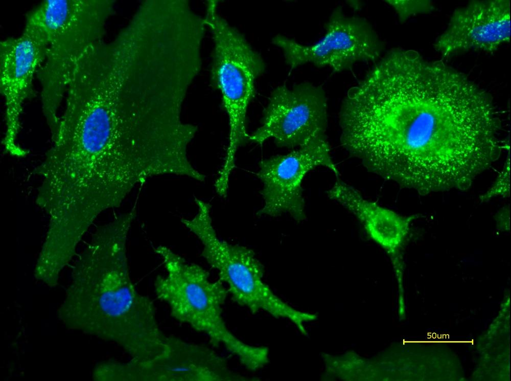
DiO membrane stained U-87 MG cells cultured on various substrates.... |  Download Scientific Diagram
DiO membrane stained U-87 MG cells cultured on various substrates.... | Download Scientific Diagram

iFluor® 488-Wheat Germ Agglutinin (WGA) Conjugate | AAT Bioquest

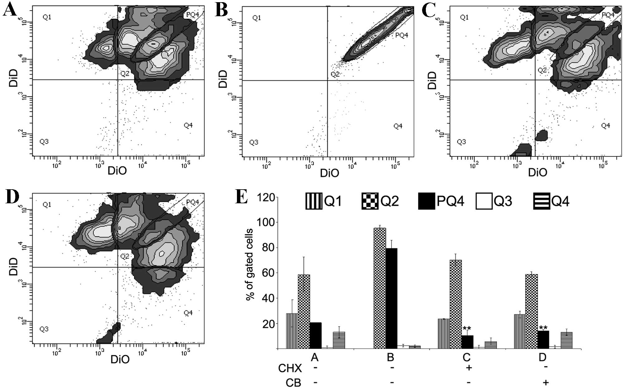
Quantification of the asymmetric migration of the lipophilic dyes, DiO and  DiD, in homotypic co-cultures of chondrosarcoma SW-1353 cells
Quantification of the asymmetric migration of the lipophilic dyes, DiO and DiD, in homotypic co-cultures of chondrosarcoma SW-1353 cells

DiO perchlorate | Cell Organelle Stains | Fluorescence Technology |  Products | MoBiTec Molecular Biotechnology
DiO perchlorate | Cell Organelle Stains | Fluorescence Technology | Products | MoBiTec Molecular Biotechnology

DiI and DIO Staining Procedures

Co-localization of Dox and CBMVs in Dox-CBMVs and on cells. a Confocal... |  Download Scientific Diagram
Co-localization of Dox and CBMVs in Dox-CBMVs and on cells. a Confocal... | Download Scientific Diagram

DiI and DIO Staining Procedures

Optimization of staining conditions for microalgae with three lipophilic  dyes to reduce precipitation and fluorescence variability - Cirulis - 2012  - Cytometry Part A - Wiley Online Library
Optimization of staining conditions for microalgae with three lipophilic dyes to reduce precipitation and fluorescence variability - Cirulis - 2012 - Cytometry Part A - Wiley Online Library

CellBrite™ Fix: Fixable membrane dyes for live cells | The Scientist  Magazine®
CellBrite™ Fix: Fixable membrane dyes for live cells | The Scientist Magazine®

Vybrant™ DiO Cell-Labeling Solution

DiI, DiD, DiR, DiO, DiA

DiO perchlorate [3,3-Dioctadecyloxacarbocyanine perchlorate] *CAS#:  34215-57-1* | AAT Bioquest
DiO perchlorate [3,3-Dioctadecyloxacarbocyanine perchlorate] *CAS#: 34215-57-1* | AAT Bioquest

A plasma membrane dye, DiO, was used to stain the histological section... |  Download Scientific Diagram
A plasma membrane dye, DiO, was used to stain the histological section... | Download Scientific Diagram

DiI and DIO Staining Procedures

DiO perchlorate | ABP Biosciences

DIO Stain Care | Chennai

DiO - Biotium
DiO - Biotium

DIO Stain Care | Chennai